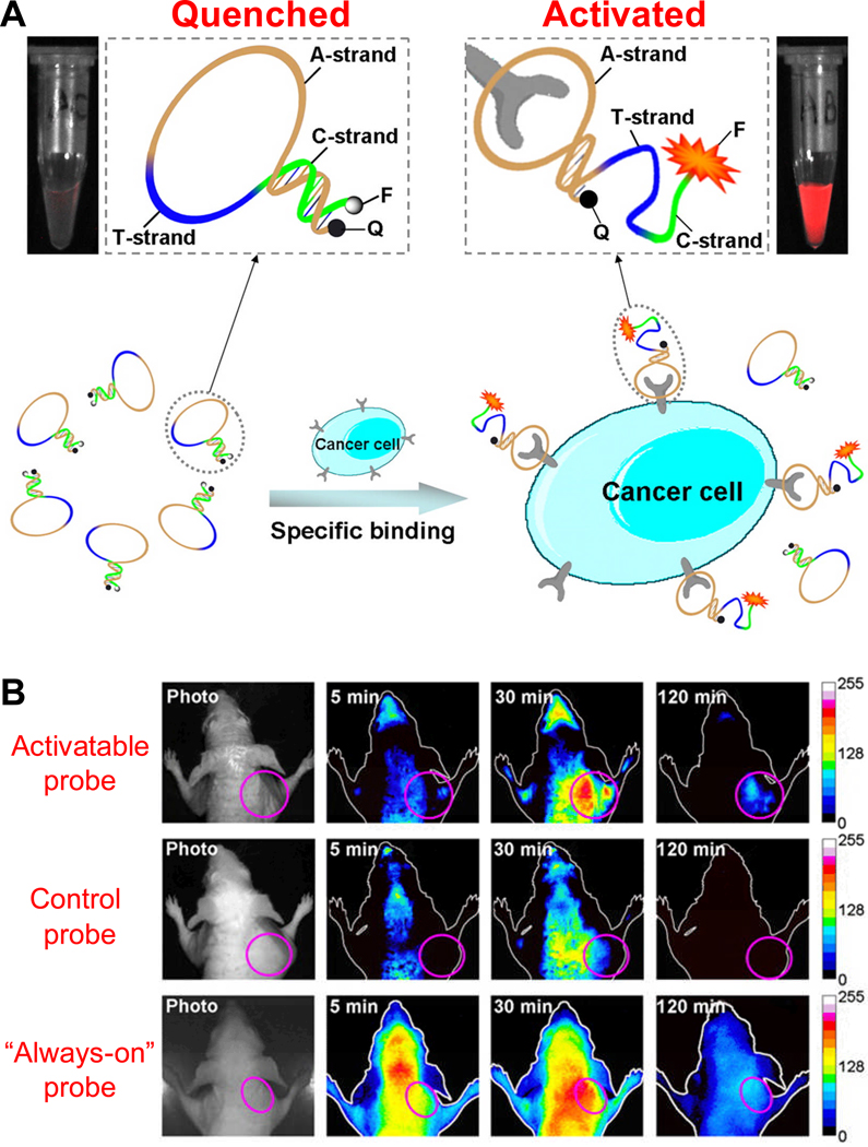
Fig. 2.
Fluorescence imaging with an aptamer-based activatable probe. A. A schematic representation of the imaging strategy based on cell membrane protein-triggered conformation alteration. The probe is consisted of three fragments: a cancer-targeted aptamer sequence (A-strand), a poly-T linker (T-strand), and a short DNA sequence (C-strand) complementary to a part of the A-strand, with a fluorophore (F) and a quencher (Q) attached at either terminus. When no target is present, the probe is hairpin-structured with quenched fluorescence. When the probe binds to targeted cancer cells, its conformation is altered which results in enhanced fluorescence signal. B. Serial in vivo fluorescence imaging of tumor-bearing mice after intravenous injection of the activatable probe, a control probe, or an “always-on” probe. The circles in the images denote the tumor sites. Adapted from [59].

