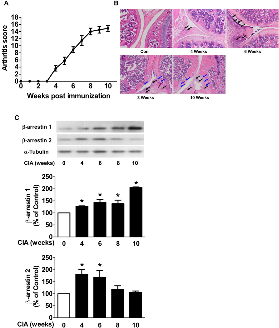
Figure 1. β-arrestin 1 and 2 expression in joint tissue in CIA mice.
CIA was studied in DBA/1J mice. The mean arthritis score reveals the disease severity (A). The hind knee joint tissue was collected from CIA mice and control mice at 4, 6, 8 and 10 weeks after the first collagen injection. Sections of joint were subjected to H&E stain (B). Black arrows indicate the PMNs and macrophages that infiltrated into synovial tissue and cavity and blue arrows indicate the bone erosion. The hind knee joints (C) were homogenized and subjected to Western blot. The densitometric levels of the scanned gels were normalized to control levels. α-Tubulin was used as an internal control. Data represent means ± S.E of five to nine mice per group from three independent experiments. *p<0.05 compared to the control mice.

