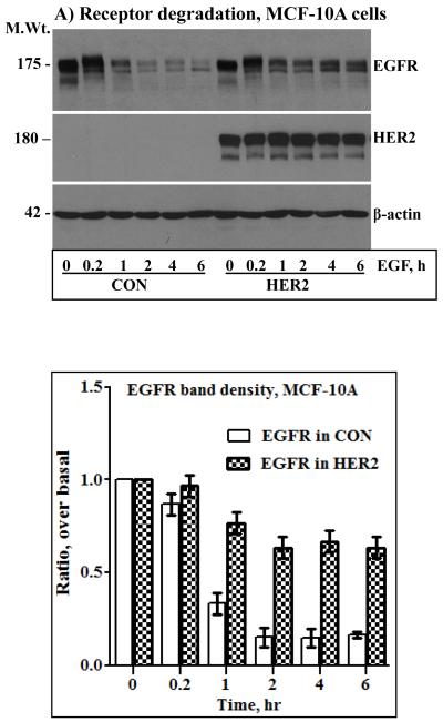
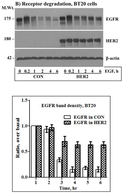
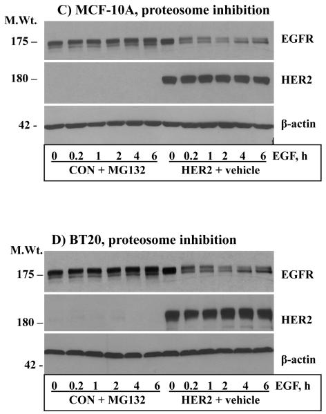
Figure 3.
Anti-HER2 and anti-EGFR immunoblotting analysis of total cell lysates prepared from the control and the HER2 cells derived from the MCF-10A (A) and the BT20 (B) lines. Cells were stimulated with EGF for the indicated time points. The bar graphs in the lower panels of A and B represent band density measurements of EGFR immunoblots presented as mean ± standard deviation from three independent experiments. Treatment of the MCF-10A (C) and the BT20 (D) cells with the general proteosome inhibitor MG-132 blocks EGF-induced EGFR degradation. Cells were treated with 5 μg/ml MG-132 for 1 hour prior to the start of EGF stimulation. Note that MG-132 completely blocks EGFR degradation while HER2 expression is relatively partial, suggesting that only heterodimerized EGFR with HER2 is protected.


