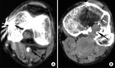
Fig. 1.
Images of arterial phase of multidetector computed tomography angiography of left lower extremity. (A) Left popliteal artery at the femoral condylar level shows an eccentric low attenuated portion, representing intraluminal thrombus (arrow). (B) At the tibial plateau level, instead of normal intravascular enhancement, a lobulated mass (empty arrows) obscured the popliteal artery and insinuated into adjacent cortex and medullary cavity of tibia resulting intraosseous mass with erosion and surrounding sclerosis (black arrow). Anteriorly, an obliquely oriented radio-dense line (white arrow) projected into the intraosseous mass was seen representing prior screw fixation site.

