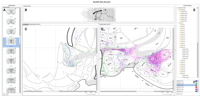
Figure 3.
A screenshot of the “standard” configuration of the NeuARt system specifically tailored to view neuroanatomical maps drawn in register with a brain atlas. This view shows detail of level 27 of (Swanson, 1998) with data maps pertaining to axonal labeling originating from the following subnuclei of the BST: oval (BSTov), fusiform (BSTfu; Dong et al., 2001b), rhomboid (BSTrh; Dong and Swanson, 2003), anterolateral (BSTal; Dong and Swanson, 2004a), and anteromedial (BSTam; Dong and Swanson, 2006a). We also include maps from PHAL injections into the lateral part of the Central nucleus of the Amygdala (CEAl) on the opposite side (Petrovich and Swanson, 1997). (A) Coronal sections for each open atlas in an “accordion” control. (B) A sagittal view of the open atlas (vertical lines are navigable). (C) Level 19 of (Swanson, 1998) in the main, vertically split central panel. (D) Level 27 of (Swanson, 1998) in the main, vertically split central panel. (E) List of the loaded data maps. (F) Control for selecting the maps to show and their color mapping.

