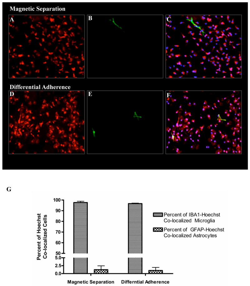
Fig. 3. Comparison of microglial cell purity obtained by differential adherence and magnetic separation.
Triple labeling immunofluorescence was with iba1/GFAP/Hoechst was used to compare microglial purity obtained by magnetic separation (A,B,C) and differential adherence (D,E,F). As shown above both methods had comparable purities with greater than 97% iba1-positive microglia. The number of Hoechst co-localized iba1-positive microglia and GFAP-positive astrocytes were also counted to quantitatively estimate the purity levels (G)

