
Figure 4.
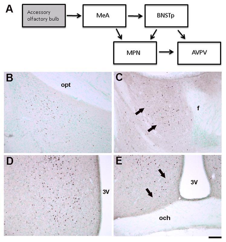
A) Schematic illustrating neural regions examined for c-Fos immunoreactivity after exposure to male-soiled bedding in the current study. The medial amygdala (MeA) receives direct projections from the accessory olfactory bulb. It projects to the BNSTp and, to a lesser extent, to the MPN. The BNSTp and MPN in turn project to the AVPV. Not all projections are shown and some, if not all, of the connections are bi-directional. B–D) Photomicrographs of c-Fos-immunoreactive cells in the MePD (A), BNSTp (B), MPN (C), and AVPV (D). 3V = 3rd ventricle; f = fornix; och = optic chiasm; opt = optic tract. Scale bar = 100 μm.