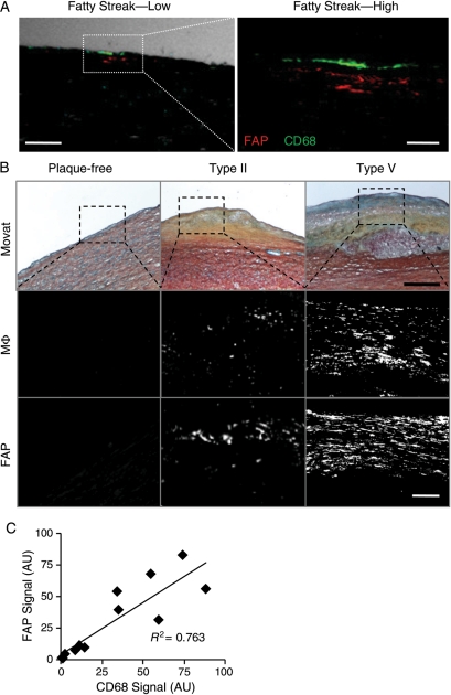
Figure 5.
Fibroblast activation protein expression correlates with macrophage burden in human aortic plaques. (A) Confocal immunofluorescent photomicrograph of an aortic fatty streak reveals fibroblast activation protein expression (red) adjacent to macrophages (CD68; green) at low (phase-contrast, white; bar = 100 μm) and high magnification (bar = 25 μm). (B) Movat staining (bar = 400 μm), fibroblast activation protein, or macrophage (CD68) immunofluorescent stainings in plaque-free aortae, type II, and type V atherosclerotic plaques show enhanced fibroblast activation protein expression with increasing macrophage burden (bar = 50 µm). (C) Comparisons of fibroblast activation protein and macrophage expression in serial adjacent sections from aortic plaques demonstrate a significant positive correlation (R2= 0.763; n = 12; P < 0.05); AU, arbitrary units.

