
Figure 5.
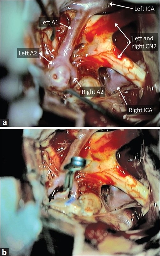
Microscopic visualization of the AComm complex through the subfrontal approach. (a) The entire AComm region and aneurysm, extending over to the contralateral side, can be easily visualized through the subfrontal approach, in this case with no gyrus rectus resection. Note that this patient did not have a right A1 segment. Note lamina terminalis (star) and perforators (arrowheads). A temporary clip is applied to the left A1 in preparation for final dissection around the aneurysm dome. (b) The view after aneurysm clipping