Figure 2.

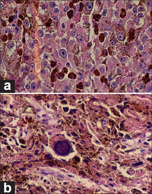
(a) Epithelioid pattern of tumor cells with dense intracytoplasmic melanin granules by H and E stain (magnification ×400). (b) Large psammoma body in the center. Large amount of melanin throughout the area of the tumor (H and E, ×400)

(a) Epithelioid pattern of tumor cells with dense intracytoplasmic melanin granules by H and E stain (magnification ×400). (b) Large psammoma body in the center. Large amount of melanin throughout the area of the tumor (H and E, ×400)