
Figure 1.
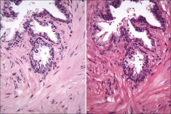
Prostate biopsy stained with the linear-batch method on the left and the HDHE method on the right. One can observe differences especially in the collagen area (screenshot of the Trestle MedMicro interface, approximate magnification 20×)
Official websites use .gov
A
.gov website belongs to an official
government organization in the United States.
Secure .gov websites use HTTPS
A lock (
) or https:// means you've safely
connected to the .gov website. Share sensitive
information only on official, secure websites.
Prostate biopsy stained with the linear-batch method on the left and the HDHE method on the right. One can observe differences especially in the collagen area (screenshot of the Trestle MedMicro interface, approximate magnification 20×)