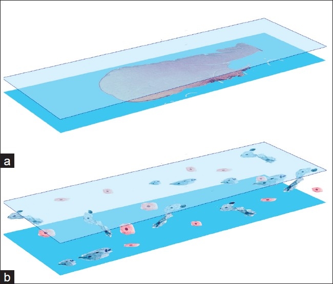
Figure 1.

Schematic illustrating differences between (a) surgical pathology slides which are usually 4-6 μm in thickness and (b) cytology slides (bottom diagram) which can range upwards of 30 μm from glass to coverslip. Cells can be positioned anywhere from glass to coverslip in cytology
