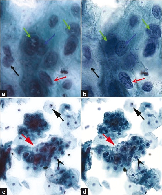
Figure 6.

Side by side comparisons between extended focus and multilayer stacking. At high power (40×), (a) extended focus improves focus (black arrow) for areas out of the plane of focus but sharpness and detail appears to be adversely affected. Nuclear contours (red arrow) and chromatin detail (blue arrow) are harder to assess, and white intranuclear digital artifacts are seen only in extended focus (green arrows). 16 focus planes were used in this example. (b) Multilayer stack counterpart to (a). At low power (10×), (c) loss of sharp detail can still be seen in extended focus compared to its multilayer stack counterpart. Nuclear detail is blurred compared to the sharply focused, corresponding area within the multilayer stack version (black arrowhead), and cell borders are difficult to assess (red arrow). (d) Multilayer stack counterpart to (c).
