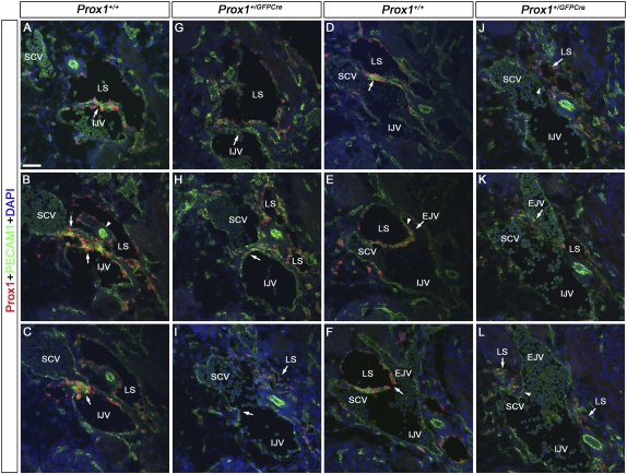
Figure 2.
Lymphovenous valves are formed by the fusion of lymph sacs with two adjacent veins. E13.5 wild-type (A–F) or Prox1+/GFPCre (G–L) embryos were transversely sectioned in an anterior to posterior orientation in the region where the jugular and subclavian veins interact (box in Fig. 1A) and were immunostained for the LEC marker Prox1 and the pan-endothelial marker PECAM1. (A) In wild-type embryos, Prox1 is expressed uniformly in LECs forming the lymph sac (LS) and in a polarized manner on the IJV (arrow). Note the relatively high levels of Prox1 on the ECs in the vein and in the lymph sacs' LECs that are facing the vein. (B) The lymph sac is split into two portions by the vertebral artery (white arrowhead). Both walls of the medial portion of the lymph sac intercalate with the wall of the IJV medially and the SCV laterally (arrows). (C) The IJV and the SCV have completely merged together, and the valve rudiment is seen in the middle (arrow). (D) The lateral portion of the lymph sac (LS) runs adjacent to the SCV. Note the relatively higher levels of Prox1 in the venous ECs and in the LECs in the lymph sac facing the vein (arrow). (E) The EJV is branching off from the SCV (arrow), and Prox1 is expressed on the walls of this vein in a polarized manner (arrowhead). This wall is also adjacent to the lymph sac. (F) The opening of the valve (arrow) is now seen and is formed by the fusion of the two layers of Prox1+ ECs. (G,H) In Prox1+/GFPCre embryos, Prox1 is expressed in LECs forming the lymph sac, but very few Prox1+ cells are seen on the walls of the IJV and SCV (arrows). (I) At the point where the IJV and the SCV merge, no Prox1+ cells or valve-like structures exist (arrow). (J) No Prox1 expression was observed on the wall of the SCV that lies close to the lymph sac (arrowhead). (K) Posterior to that, the EJV branches off from the SCV. No Prox1+ cells are seen on the walls of the EJV (arrow). (L) No communication is observed between the lymph sac and the veins (arrowhead). Also, note an overall reduction in the number of LECs in G–L. The neural tube is oriented toward the right, the heart is oriented toward the left, and the thymus is oriented toward the bottom in each panel. Bar, 50 μm.

