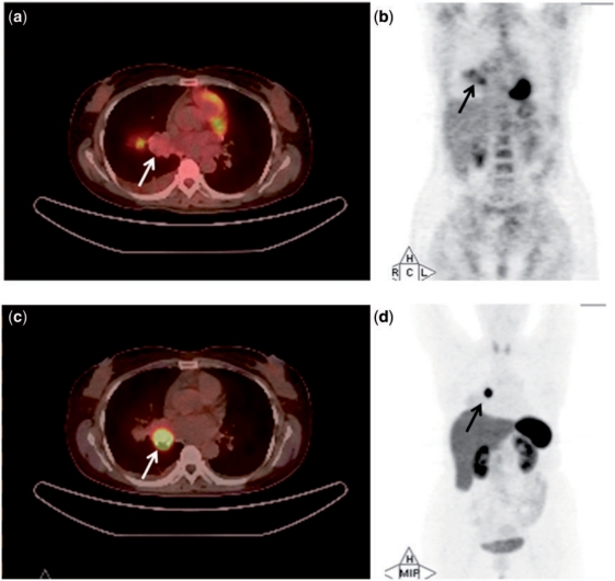
Figure 1.
PET/CT (a) and whole-body projection images (b) of a 40-year-old woman (case 1) with typical carcinoid (arrow) revealing low tumour uptake on [18F]FDG-PET/CT (SUVmax, 1.3). PET/CT image (c) and whole-body projection image (d) of the same patient revealing high tumour uptake on [68Ga]DOTATOC-PET/CT (SUVmax, 23.2).

