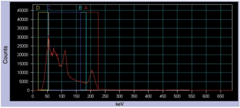
Figure 1.
Energy distribution of incident photons (spectrum) when scanning a patient injected with [177Lu]octreotate (anterior abdominal view). A is the photopeak energy window (208 keV; 20% width), B is the lower scatter energy window (10% width), C and D are general scatter windows (110.9 and 37 keV, respectively; 100% width).

