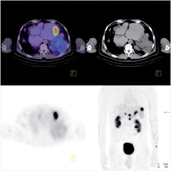
Figure 5.
From left to right, top to bottom, respectively: fused, CT and SPECT transaxial slice, and anterior maximum intensity projection of body QSPECT/CT started 60 min after intravenous administration of 8.4 GBq of [177Lu]octreotate, in a patient with metastatic neuroendocrine tumour to liver and abdominal lymph nodes (patient 1). The upper threshold of the colour scale (red) was set to SUV 12.

