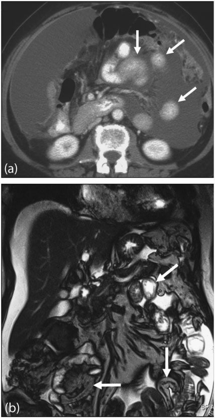
Figure 13.
Serosal deposits. (a) Axial contrast-enhanced MDCT shows small bowel serosal deposits from metastatic ovarian carcinoma (arrows). Note involvement of the greater omentum and extensive ascites. In a different case, (b) coronal T2-weighted MRI demonstrates multisegment small bowel serosal deposits (arrows).

