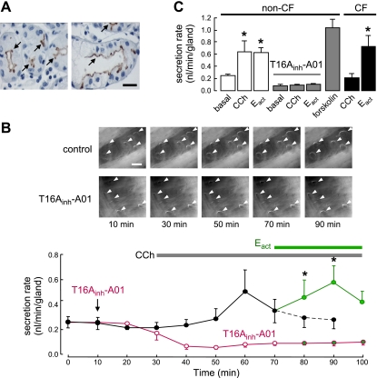
Figure 7.
Airway submucosal gland fluid secretion in human bronchi. A) TMEM16A immunohistochemistry in CF (left panel) and non-CF (right panel) human bronchi showing apical membrane expression in serous gland epithelial cells (arrows). Scale bar = 20 μm. B) Mucus (fluid) secretion in human bronchi. Top panels: images of mucus bubbles formed under oil in response to basolateral application of 300 nM carbachol (CCh) and 20 μM Eact. TMEM16A was inhibited by 30 μM T16Ainh-A01. Individual fluid bubbles marked with arrowheads. Scale bar = 0.5 mm. Bottom panels: CCh and Eact-induced secretion rates. Where indicated, tissues were pretreated with T16Ainh-A01 (30 μM). Each point is the average of measurements made from 20 glands (mean±se). C) Summary of human gland fluid secretion rates measured at 20 min after addition of 20 μM Eact, and 30 min after application of 300 nM carbachol (CCh) and 10 μM forskolin (20–66 glands from 3 tracheas and 4 bronchi). In CF-bronchi, 6 glands from one donor were stimulated by Eact. *P < 0.05.

