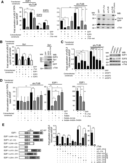
Figure 3.
γ-Tubulin moderates E2F1, E2F2, and E2F3 transcriptional activity. Assay of the luciferase activity driven by 6 E2F promoter binding sites (A, C, E) or cyclin E promoter (B, D) on transient transfection of U2OS cells with a Renilla reporter construct and the following constructs: GFP, γ-TUBULIN-shRNA (shγTub), HA-E2F1, HA-E2F2, HA-E2F3, HA-E2F6, HA-Dp1, GFP-γ-tubulin (γTub), RNAi-resistant γ-TUBULIN gene (γTub-rest), E2F1-shRNA (shE2F1), E2F2-shRNA (shE2F2), E2F3-shRNA (shE2F3), Ala399-γtubGFP (R399A), Ala409-Ala410-γtubGFP (R409A-K410A), Ala399-Ala400-Ala409-γtubGFP (R399A-K400A-R409A), N-γtubGFP1–333 (N-γTub), and C-γtubGFP334–452 (C-γTub), or various E2F1 mutants: HA-E2F1Δ409–426 (Δ409–426), HA-E2F1Δ2–126, HA-E2F1Δ360–426, HA-E2F1Δ193–426, Flag-E2F1Δ193–284, Flag-E2F1Δ193–359 or Flag-E2F1Δ284–359, as indicated. Luciferase activity of cells transfected with control construct was set as 1, and relative activities were calculated (means±sd, n=3–10). A–C) Total lysates of transfected U2OS cells were analyzed by WB with the indicated antibodies. Arrowheads and arrows indicate GFP-γ-tubulin and endogenous γ-tubulin, respectively. E) Structure of wild-type E2F1 and various E2F1 constructs comprising the DNA-binding (DBD), dimerization (DD), transactivating (TAD), RB-binding (RBD), and γ-tubulin-interacting (γD) domains. *P < 0.05; **P < 0.01.

