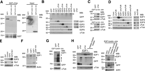
Figure 4.
γ-Tubulin affects the expression levels of E2F1, E2F2, and E2F3. A) Purified full-length GST-γ-tubulin was incubated with His-E2F1, His-E2F1Δ349–426, or His-E2F1Δ193–426, and the GST-γ-tubulin was subsequently retrieved by adsorption to glutathione-Sepharose 4B and examined by WB (n=3). Total amounts of loaded His-tagged proteins were analyzed by WB (input). B–H) Total lysates of transfected U2OS cells with the following constructs: GFP, HA-E2F1, -E2F2, -E2F3, -E2F6, -Dp1, GFP-γ-tubulin (γTub), γ-TUBULIN-shRNA (shγTub), HA-E2F1Δ409–426 (Δ409–426), Flag-E2F1Δ193–284, Flag-E2F1Δ193–359, Ala399-Ala400-Ala409-γtubGFP (R399A-K400A-R409A), γtubGFP-R409A-K410A (R409A-K410AGFP), or empty vector (Cont.) were analyzed by WB with the indicated antibodies. Arrows and arrowheads indicate GFP-fused proteins and E2F1 degradation products, respectively (n=3). D) Overexposed WBs of the endogenous expression of E2Fs are shown at right.

