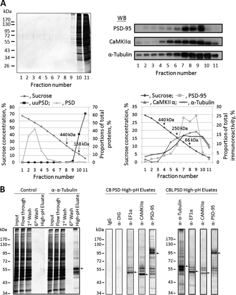
Fig. 6.
Immunoabsorption of PSD proteins. A, Sucrose-density-gradient centrifugation analysis of the uuPSD and original PSD samples isolated from cerebral cortex. Top left panel: SDS-PAGE analysis of the fractions collected from the 0–70% gradient of uuPSD (0.2 mg protein) as visualized by silver staining. The molecular weight markers are shown to the left. Bottom left panel: Protein contents of the fractions collected from the gradients of the uuPSD (■) and PSD (▴) samples. The protein content of each fraction is expressed as the percentage proportion of the fraction's integrated protein intensity against the sum of the integrated intensities of all collected fractions as calculated from the densitometric scan of the gel after silver staining. The positions of two standards, ferritin (440 kDa) and aldolase (158 kDa) are indicated by arrows. Top right panel: The high-pH-eluates collected from immunoabsorption experiments using the antibodies against PSD-95, CaMKIIα and α-tubulin were subject to sucrose-density-gradient centrifugation analysis, and the collected fractions were subject to Western blotting analysis by using antibodies against PSD-95, CaMKIIα and α-tubulin. Bottom right panel: Quantification of the intensities of the inmmunostained bands found in the top panel. The intensity of an immunostained band in a fraction is expressed as the proportion of its intensity against the sum of the intensities of all fractions. The positions of ferritin (440 kDa), catalase (250 kDa), and bovine serum albumin (66 kDa) are indicated by arrows. B, SDS-PAGE analyses of the fractions collected in immunoabsorption experiments. Left panel: Aliquots of 38 μl were removed from the indicated fractions of the immunoabsorption experiments conducted with cerebral PSD wherein no antibody (control) or the anti-α-tubulin antibody was used for SDS-PAGE analyses. Molecular weight markers are shown to the left. Middle and right panels: Aliquots of 38 μl were removed from the high-pH-eluates obtained from the immunoabsorption experiments of cerebral (CB) and cerebellar (CBL) PSDs, respectively, using mouse idiotypic monoclonal antibody (IgG) and antibodies against DIG, EF1α, CaMKIIα, and PSD-95 and subject to SDS-PAGE analysis. Arrows indicate the major protein bands found in the high-pH-eluates, which are of the same sizes as the antigens of the antibodies used in the immunoabsorption experiments. The results are from a representative experiment out of a total of 3 to 6 experiments.

