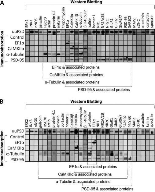
Fig. 7.
Western blotting analyses of proteins co-immunoabsorbed with EF1α, CaMKIIα, α-tubulin and PSD-95 in cerebral PSD (A) and cerebellar PSD (B). The uuPSD (10 μg protein) and the high-pH-eluates obtained in the immunoabsorption experiments using no antibody (control) or the antibodies against EF1α, CaMKIIα, α-tubulin and PSD-95 (1/8 of the collected samples) were subject to Western blotting analysis by using antibodies against 29 PSD proteins. The gels shown are from a representative experiment out of at least three independent experiments. Asterisks indicate the immunostained bands that colocalize with the major proteins found in the high-pH-eluates of immunoabsorption experiments (as indicated by arrows in Fig. 6B) by using the same antibodies. Proteins associated to the above antigens are indicated by the brackets at the bottom.

