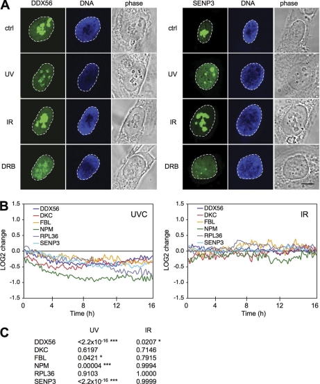
Fig. 2.
Fluorescent-tagged nucleolar proteins relocalize in a damage-specific manner. A, U2OS cells stably expressing DDX56-GFP or SENP3-YFP fusion proteins were treated with UV (35 J/m2), DRB (100 μm), or IR (10 Gy) for 6 h or left untreated and fixed. Nuclei were counterstained with Hoechst 33258. Confocal images are shown. Scale bar, 10 μm. B, Live-cell image analyses of U2OS cells stably expressing YFP/CFP-tagged proteins. Cells were treated with UV (35 J/m2) or IR (10 Gy) or left untreated and stained with Hoechst 33342 (1 μg/ml). Cells were imaged with Stallion HSI wide-field microscope for 16 h and images were captured every 10 mins. Individual cells were tracked, and nucleolar and nucleoplasmic intensities were recorded. Intensity difference between nucleoli and nucleoplasm for individual cells (n = 40–200) was analyzed, normalized to untreated cells and plotted as LOG2 values. C, p values were calculated by Two-Way ANOVA analysis.

