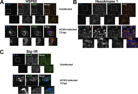
Fig. 8.
Relocalization of Sig-1R during HCMV infection of HFFs. HFFs were uninfected or HCMV infected at a multiplicity of 1 and harvested at 72 hpi. A, Induction of HSP60 during HCMV infection. Uninfected and HCMV-infected HFFs were probed with anti-HSP60 (green), anti-HCMV IE 1/2 (blue), and human anti mitochondria (red) antibodies and the corresponding secondary antibodies. The stained cells were imaged by confocal microscopy. The three leftmost panels are greyscale and the right panel shows the overlay of confocal sections. The insets show enlarged regions of interest in the cells. B, Induction of Hexokinase 1 at late times of HCMV infection. Cells were probed with anti-Hexokinase (green), anti-HCMV IE 1/2 (blue), and human anti mitochondria (red) and the corresponding secondary antibodies and imaged as above. The three leftmost panels are greyscale and the right panel shows the overlay of confocal sections. The insets show enlarged regions of interest in the cells. C, Relocalization of Sig-1R during HCMV infection of HFFs. Cells were probed with anti-Sig 1R (green) and anti HCMV IE 1/2 (blue) antibodies. The left and middle panels are greyscale and the right shows the overlay of confocal sections. The insets show enlarged regions of interest in the cells.

