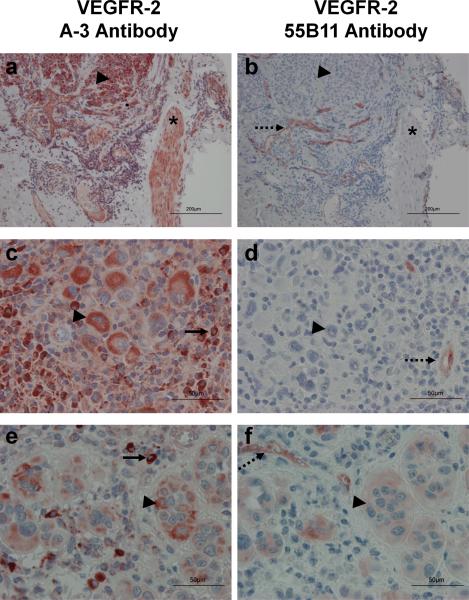
Figure 1.
Immunohistochemistry of melanoma TMAs. TMAs were processed and stained as described under materials and methods with antibody A-3 (panels a, c, and e), or antibody 55B11 (panels b, d, and f). Endothelial cells serve as an internal positive control for VEGFR-2 (dashed arrows). Smooth muscle cells (*), histiocytes (solid arrows), and melanoma cells (arrowheads) are indicated. (Magnification, a and b: X100, scale bar = 200 um; c, d, e and f: X400 and scale bar = 50 um).

