Figure 4.
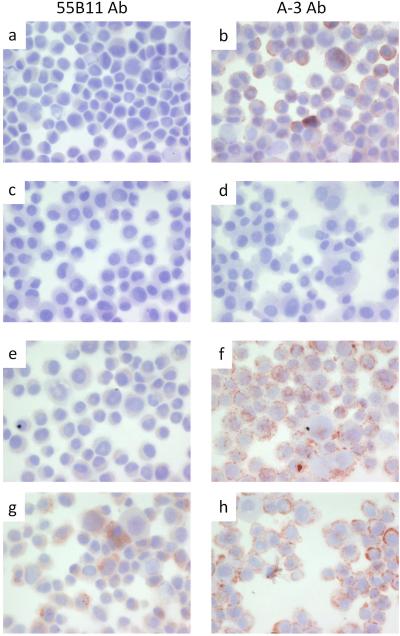
Immunohistochemistry of DM122 melanoma cells. Cytospins were processed and stained as described under materials and methods with antibody 55B11 (panels a, c, e, and g), or antibody A-3 (panels b, d, f, and h). DM122 melanoma cells alone (positive control) are shown in panels a and b. DM122 melanoma cells without primary antibody (negative control) are shown in panels c and d. DM122 melanoma cells mock transfected are shown in panels e and f. DM122 melanoma cells transfected with plasmid encoding VEGFR-2 are shown in panels g and h.

