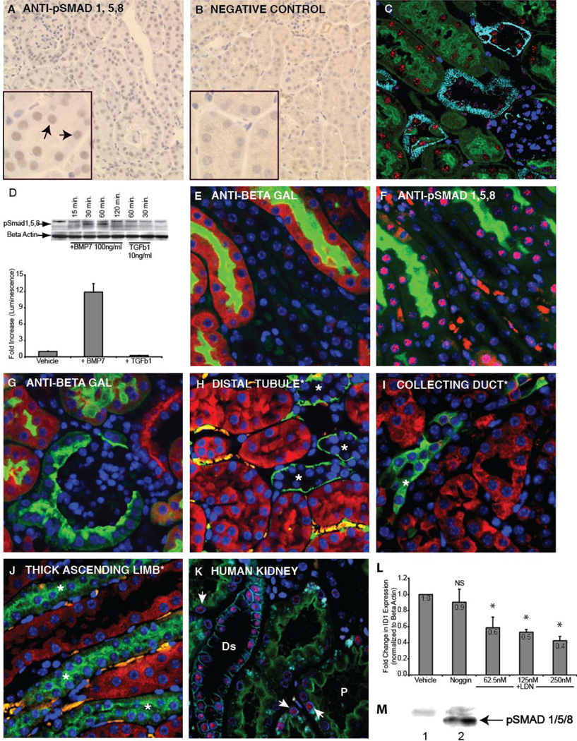
Figure 1. BMP signaling in the adult mouse and human kidney.
A: Immunohistochemical staining for SMAD 1/5/8 (Cell Signaling Technology) reveals nuclear accumulation in mouse nephron epithelia. B: In the negative control, the primary antibody was omitted. C: Immunofluorescent staining of mouse kidney using monoclonal antibody against pSMAD1/5/8 (clone Vli-49), red), antibody against E-cadherin marking distal tubule (light blue), Lotus lectin marking proximal tubule (green) and DAPI (dark blue). D: Immunoblot comparing SMAD1/5/8 phosphorylation following BMP7 or TGFβ1 treatment of human primary renal proximal tubule epithelial cells (top panel). pBRE-Luc BMP transcriptional reporter assay in HK-2 human proximal tubule cells stimulated either with BMP7 (50 ng/ml) or TGFβ1 (5ng/ml) for 18 hours. E: Immunofluorescent staining of the reporter mouse kidney using an antibody against β-galactosidase (red) indicates that the highest level of BMP signaling is within the proximal tubule (green, lotus lectin). F: An adjacent section stained with a monoclonal anti pSMAD1/5/8 (Vli-49, red) shows pathway activation in a pattern consistent with reporter activation. Lotus lectin (green) labels proximal tubules. G: Staining for β-galactosidase (red) shows absence of staining in the glomerulus. Panels H, I, and J show BMP signaling (red) relative to molecular markers. H: Distal tubule (green, anti-E-cadherin). I: Collecting duct (green, DBA lectin). J: Thick ascending limb of the distal tubule (green, anti-Tamm Horsfall protein). K: In the human kidney, BMP signaling (red, monoclonal anti-pSMAD1/5/8 Vli-49) is most intense in the distal tubule (light blue, anti-E-cadherin), and weaker signal is seen in the proximal tubule (green, Lotus Lectin, arrows). L: Expression of ID1 was measured in RPTECs incubated for 14 hours with the BMP signaling inhibitor, LDN-193189. To control for BMP in medium, Noggin was added. Asterisk indicates a p value (relative to vehicle) of less than .05. M: Western blot analysis on MLFM4 cell lysate using rabbit monoclonal (Vli-49) anti-pSmad 1/5/8: Lane 1, untreated, lane 2 treated with BMP7 (50ng/ml). Abbreviations: Ds, distal tubule; P, proximal tubule, NS, not significant (p value greater than .05).

