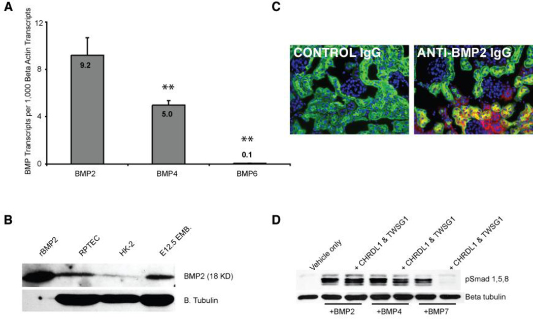
Figure 2. BMP is expressed in the proximal tubule.
A: QPCR showing the number of BMP transcripts per 1,000 copies of β-actin transcripts. Double asterisks indicate a p value of less than .01 (when compared to BMP2 expression). B: Immunoblot showing BMP2 expression in RPTECs. Recombinant BMP2 protein and lysate from an E12.5 embryo were used as positive controls. C: Immunofluorescent micrographs of the kidney showing BMP2 (red) expression in renal epithelia, including proximal tubules (green, lotus lectin). In the left panel, nonspecific IgG was used as a negative control. D: The proximal tubule expressed BMP antagonist CHRDL1 antagonizes BMP7 but not BMP2 or 4 in conjunction with Twisted gastrulation (TWSG1). RPTECs were serum starved for 4 hours and incubated with BMPs (50ng/ml) and antagonists (400 ng/ml) for 30 minutes prior to lysis and western blotting for pSMAD1/5/8.

