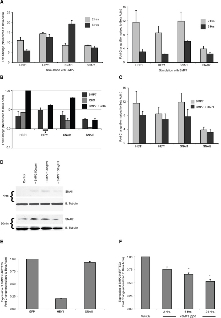
Figure 4. BMP signaling directly activates a set of typical Notch response genes.
A: QPCR confirming that HES1, HEY1, SNAI1 and SNAI2 are strongly up-regulated in RPTECs after treatment for 2 or 6 hours with 50ng/ml of BMP2 (left panel) and BMP7 (right panel). The less durable response of BMP7 at 6 hours may result from increased amounts of secreted CHRDL1 accumulating in the medium and resulting in greater antagonism of BMP7, but not BMP2. B: RPTECs were incubated for 30 minutes with cycloheximide (5µM) or vehicle prior to the addition of BMP7 (50ng/ml) for 2 hours and analyzed by RT-qPCR. Cycloheximide did not reduce target gene activation showing that protein synthesis is not required for BMP7 induction of expression. C: Cells were incubated with vehicle or 5µM DAPT for 30 minutes prior to adding BMP7 at 50ng/ml. qPCR shows that the inhibition of Notch signaling did not significantly reduce gene induction at two hours. D: REPTCs were serum starved for 2 hours, then treated for the time specified with BMP2 at 50 and 100ng/ml or with BMP7 at 100ng/ml. Immunoblots were probed for SNAI1 (Snail) and SNAI2 (Slug). Blots for Beta tubulin below each protein of interest show relative loading of sample. E: qPCR showing BMP2 expression in RPTECs transduced for 32 hours with adenoviral vectors expressing GFP (control), HEY1 or SNAI1. F: qPCR showing reduction in BMP2 expression in RPTECs incubated with 50ng/ml of BMP2 for 2, 6, and 24 hours. Asterisk indicates p value < 0.05.

