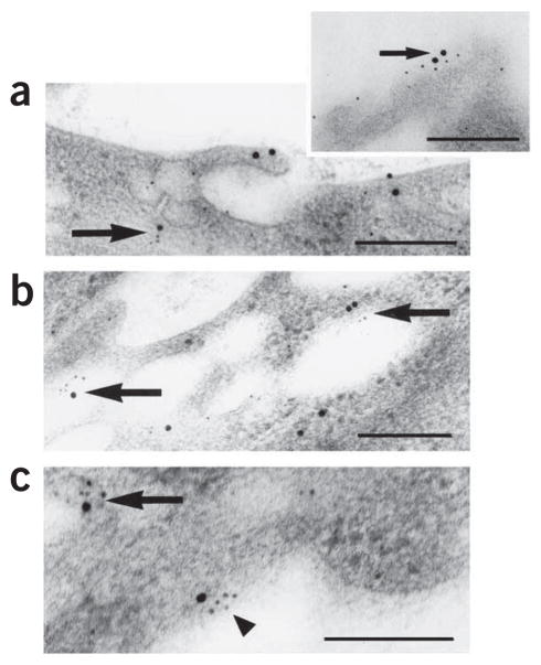
Figure 1.
Colocalization of DARC and CXCL8 in venular endothelial cells in human skin. Ultrastructural colocalization of DARC immunoreactivity (large black dots; silver-enhanced 15-nm colloidal gold) with CXCL8 immunoreactivity (small black dots; silver-enhanced 5-nm colloidal gold) in plasmalemmal vesicles (a) and on the luminal membrane (inset, a), in large intracellular electron-lucent vesicles (b) and on the abluminal membrane (c) in venular endothelial cells 120 min after the injection of CXCL8 (12 pmol) into intact pieces of normal human skin immediately after removal during elective surgery. Large arrows, small arrow and arrowhead indicate colocalization intracellularly, luminally and abluminally, respectively. Scale bars, 200 nm. Results are representative of two experiments with skin samples from two people.

