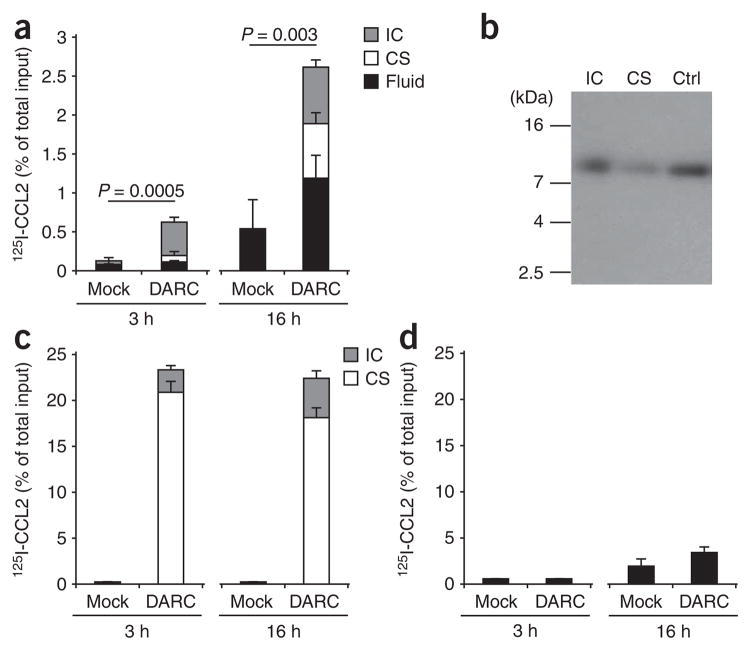
Figure 3.
Chemokine transport by DARC across MDCK-DARC monolayers. Analysis of 125I-labeled CCL2 (125I-CCL2) placed above or below monolayers of MDCK-mock cells (Mock) and MDCK-DARC cells (DARC) and, after incubation, detected in three different compartments: intracellular (IC), cell surface bound (CS) and in the fluid (Fluid). (a) Basolateral-to-apical transport of intact 125I-labeled CCL2 TCA-precipitable. (b) Autoradiography of MDCK-DARC cell–associated 125I-labeled CCL2 after its basolateral-to-apical transport. Ctrl, native 125I-labeled CCL2 (control). kDa, molecular size in kilodaltons. (c,d) Binding and internalization (c) and transport (d) of intact 125I-labeled CCL2 from the apical side to the basolateral side. (d) Soluble chemokine in the bottom Transwell chamber. Data are representative of three (a,c,d) or two (b) independent experiments (mean and s.d. of three samples).

