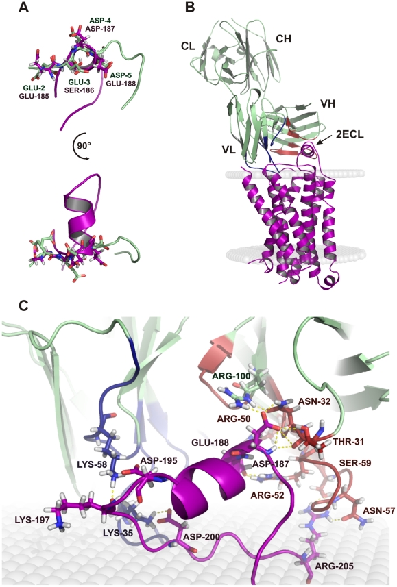
Figure 4. Model of the interaction of Fab 17.2 with the human β1-AR.
A. Superposition of the second extracellular loop (2ECL) of the human β1-AR model (violet) and the region 2–5 of the epitope (light green). B. Complex of the Fab 17.2 (light green) with the human β1-AR (violet) inserted in a membrane model (grey). C. Zoom of the paratope region of 17.2 interacting with the second extracellular loop. The main contact points are illustrated as dotted yellow lines. Heavy chain CDRs are coloured in red and light chain CDRs in blue.

