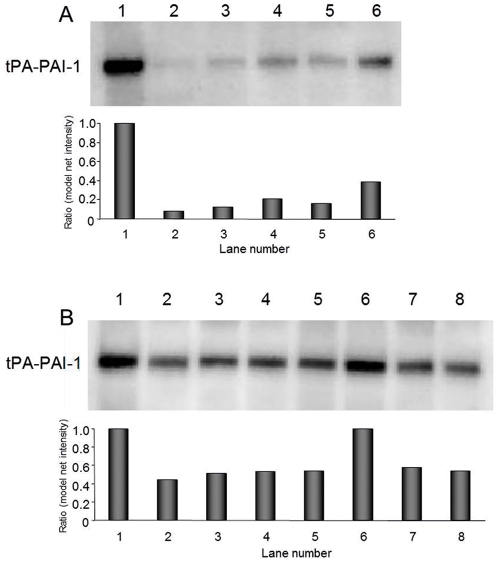
Figure 4. Western blot analysis of tPA-PAI-1 complex showing the differences in PAI-1 activity depending on the lysis method.
In all samples, equal number of platelets (180×106) was lysed and equal amounts of tPA (500 ng) was used. The formation of tPA-PAI-1 complex was detected by the PAI-1 mab (MAI-12). Each lane represents different lysis conditions. Sonicated samples on membrane A was lysed with high energy on setting 7. On membrane B, sonicated samples were lysed with milder sonication on setting 2. To lane 1, 5, 6, and 7 tPA was present during lysis. In lane 2, 3, and 4 tPA was added to the samples after lysis. Lysis conditions of the platelets: Lane 1A, 1B: in lysis buffer containing 0.1% Triton X-100 with tPA present. Lane 2A, 2B: in homogenisation buffer lysed by sonication. Lane 3A, 3B: in Pipes buffer lysed by sonication. Lane 4A, 4B: in Pipes buffer lysed by freezing and thawing. Lane 5A, 5B: in Pipes buffer lysed by sonication with tPA present. Lane 6A: in Pipes lysed by freezing and thawing with tPA present. Lane 6B 7B and 8B: 6 same as sample 1, 7 and 8 same as 3 and 4, but with 0.1% Triton X-100 added after sonication or freezing/thawing. The results of the densitometry measurements are presented as arbitrary units with samples lysed by Triton X-100 set to 1.0.

