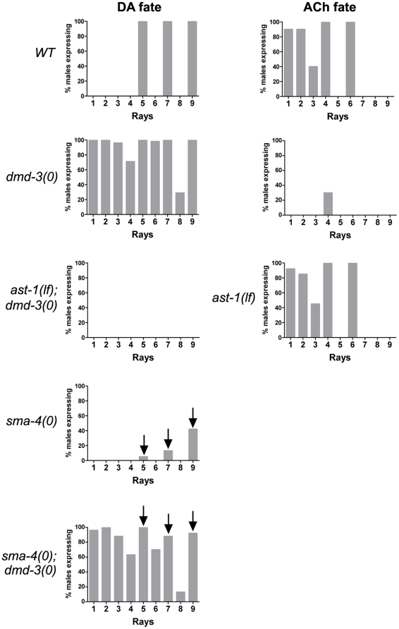
Figure 3. Quantification of ray neurotransmitter patterning defects in dmd-3, ast-1 and DBL-1 pathway mutants.
Populations of males of the genotype shown and carrying DA or ACh fate reporter transgenes were scored for the frequency of marker expression in rays indicated on the X-axis. The Y-axis shows the percentage of males that express the marker in a particular ray. n = 40–100 male tail sides scored (see Tables S1 and S2 for additional data). Except for ast-1 mutants, the DA fate marker is pdat-1::GFP. For ACh fate, unc-17::GFP marker expression is shown. In wild type (WT) males, DA fate is restricted to the A-neurons of rays 5, 7 and 9 and ACh fate is expressed in the A-neurons of rays 1 to 4 and 6. In dmd-3(0) males, the A-neurons of additional rays variably express DA fate while ACh fate is almost completely eliminated. In dmd-3(0) males, the frequency of DA fate expression (visualized with CAT-2::GFP) is significantly reduced by ast-1 loss of function. ast-1 loss of function has no effect on ACh fate expression. sma-4 encodes a SMAD of the DBL-1 signal transduction pathway that promotes rays 5, 7 and 9 identity. In sma-4(0) males, the A-neurons of these rays adopt DA fate at low frequency. Removal of dmd-3 activity in a sma-4(0) background restores DA fate expression to 100% in these A-neurons (indicated by the arrows).

