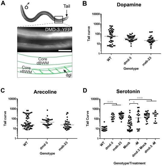
Figure 6. dmd-3 and mab-23 mutants have functionally abnormal muscles.
A. DMD-3::YFP is expressed in core and male-specific muscles. A fluorescent micrograph showing DMD-3::YFP expression in muscles of the male posterior (lateral view, dorsal uppermost, posterior to the right). The region shown corresponds to the boxed area on the worm image above. DMD-3::YFP is expressed in dorsal and ventral core body wall muscles (dBWMs and vBWMs, respectively) and in the male-specific diagonal muscles (dgl, the arrow indicates a single dgl muscle). The dgl muscles lie immediately beneath the core vBWMs. Magnification: 600×. Scale Bar = 10 µM. B–D. Tail curves generated by adult males after exposure to exogenous neurotransmitter receptor agonists. Adult males of the genotype or treatment shown (X-axis) were incubated in solutions of B. 50 mM dopamine C. 1 mM arecoline D. 50 mM serotonin and their tail curls were measured (Y-axis log scale; see Materials and Methods). Each dot corresponds to the performance of a single male. In arecoline, wild type males can adopt a dorsal (grey squares) or ventral curl (black dots). In dmd-3 and mab-23 mutant populations, dorsal curls are infrequent, consequently the ratio of dorsal: ventral tail curls for the two populations is significantly different from that of wild type (p<0.001). However the form of the ventral curl posture produced by mutants is similar to that of wild type males (see Fig. S2). In D, the “-M” treatment corresponds to males in which the M sex-muscle precursor cell has been ablated. The “Mock” treatment corresponds to males which were exposed to anesthetic but not subjected to laser ablation treatment. Results of Nonparametric 1-way ANOVA (Kruskal-Wallis) are shown. Median, grey bars. Significance *** p<0.001; ** p<0.01; * p<0.05.

