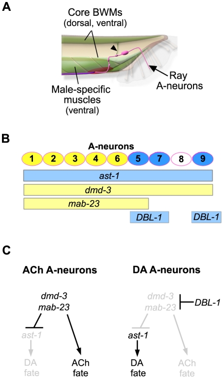
Figure 7. dmd-3 and mab-23 specify multiple cell fates in the ray sensorimotor circuit.
A. Schematic of the male tail (lateral view, posterior right) indicating those tissues that express dmd-3 and mab-23 reporters. In the A-neurons of the rays (pink), mab-23 and dmd-3 regulate DA and ACh fates. In the muscles (green), dmd-3 and mab-23 regulate unknown characteristics that render the muscles responsive to ray circuit control. Ray axons synapse directly on to muscles of the tail en route to the ventral pre-anal ganglion (red oval). Synapses with dorsal muscles occur on muscle arms that project ventrally (arrowhead). The rays also make indirect connections with muscles through inter- and motor neurons of the pre-anal ganglion and ventral nerve cord (purple line). B. Sites of action for ast-1, dmd-3, mab-23 and the DBL-1 pathway in the A-neuron population. The A-neurons (circles) of rays 1 to 9 stereotypically express ACh (yellow) or DA (blue) fate. The bars below indicate the zone of activity for a specific regulator within the A-neuron population. ast-1 and the DBL-1 pathway (blue bars) promote DA fate while dmd-3 and mab-23 (yellow bars) repress DA fate and promote ACh fate. C. A genetic model for control of A-neuron neurotransmitter fate. In the A-neurons of rays 1 to 4 and ray 6, dmd-3 and mab-23 activity promote adoption of ACh fate and simultaneously suppress DA fate, which is conferred by ast-1. The A-neurons of rays 5, 7 and 9 are differentially exposed to the DBL-1 ligand during development. Activation of the DBL-1 signal transduction pathway within the A-neurons of these rays blocks dmd-3 and mab-23 activity. Consequently, ACh fate is not expressed and ast-1 can activate transcription of dopamine terminal fate genes.

