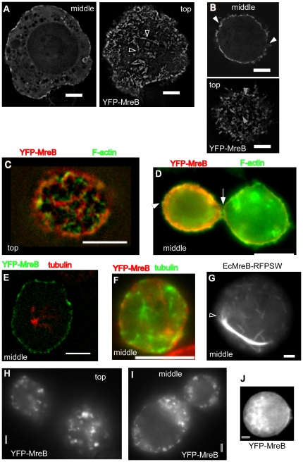
Figure 1. Expression of YFP-MreB or mutant versions in S2 cells.
A) Wild type YFP-MreB filaments, shown are a middle plane and top plane of a Z-stack. Triangles indicate bundles of filaments from which a single filament (or thin bundle of filaments) emanates. B) Middle and top planes of a 3D deconvoluted Z-stack of a cell expressing wild type YFP-MreB. C–D) Immunofluorescence of cell expressing YFP-MreB, using phalloidin as stain for actin filaments. Triangles indicate positions of actin filaments that lack any detectable YFP-MreB fluorescence. E–F) Immunofluorescence of cell expressing YFP-MreB, using anti Drosophila tubulin antiserum to stain for tubulin filaments. G) E. coli MreB (with an internal RFP) expressed in S2 cells, shown is the middle plane. Triangle indicates MreB filaments extending from the end of a filament bundle. H–I) Cells were depleted for ATP by the addition of FCCP, H–I) 20 min after addition, J) 90 min after addition (middle plane is shown). White bars 2 µm (A,B, G) or 5 µm (C–F) respectively, grey bars 2 µm.

