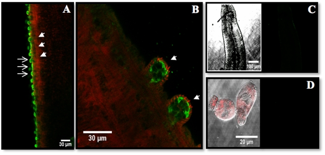
Figure 5. Tissue localization of SmGBP.
S. mansoni were probed with affinity-purified anti-SmGBP antibody followed by a FITC-labeled secondary antibody. (A) Strong immunoreactivity (green) was detected on the surface layer of adult male worms (open arrows). Actin-rich muscles were stained with TRITC-labeled phalloidin (red) and can be seen beneath the tegument (solid arrow heads). At higher magnification, the SmGBP signal was found to be enriched in the tubercles (arrows) of the male, where numerous actin-rich phalloidin-labeled spines (red) can also be seen (B). No significant SmGBP immunofluorescence was detected in male worms probed with peptide-preadsorbed antibody (C). Besides males, we did not observe specific SmGBP immunoreactivity in any of the other stages tested, including cercariae (D) schistosomula and female worms (not shown).

