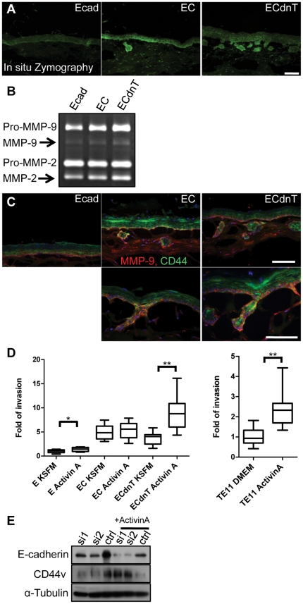
Figure 4. CD44 co-localizes with MMP-9 in areas of invasion and is upregulated by Activin A.
(A) For in situ zymography, areas of MMP activity are highlighted by a positive fluorescein signal on frozen sections of Ecad, EC and ECdnT organotypic cultures. (B) Zymography using organotypic culture conditioned medium collected from Ecad, EC and ECdnT cells illustrates increased secretion of MMP-2 and MMP-9. (C) Double immunofluorescence staining of Ecad, EC and ECdnT shows MMP-9 (red) colocalization with CD44 (green) in invasive cells. Scale bar is 50 micron. (D) Stimulation with Activin A induces enhanced invasion of ECdnT and TE-11 cells in invasion assays. Comparison between groups was done by Man and Whitney non- parametrical test, * p<0.05, **p<0.001. (E) Western Blot of cell lysates from TE-11 transfected with AllStars negative non-silencing siRNA cells (ctrl), and TE-11 cells transfected with siRNA against E-cadherin (si1, si2) demonstrates upregulation of CD44 after stimulation with Activin A.

