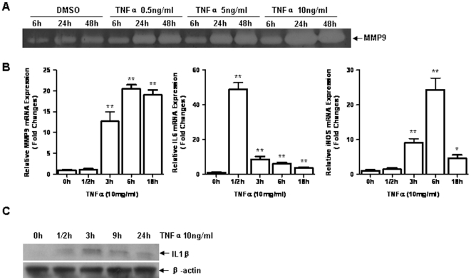
Figure 1. TNF-α induced overexpression of MMP-9, iNOS, IL-6 and IL-1β in 3T3/NIH fibroblasts. A.
3T3/NIH cells were incubated for 6, 24 or 48 hours (h) with varying concentrations of TNF-α, gelatin zymography were performed to detect MMP-9 expression in the medium. B. Cells were treated by TNF-α (10 ng/ml) for 0, 1/2, 3, 6, 18 h, relative mRNA expressions of MMP-9, iNOS and IL-6 were examined by real-time RT-PCR analysis (n = 3 per group, *p<0.05 vs. 0 h; **p<0.01 vs. 0 h). C. Cells were treated by TNF-α for 0, 1/2, 3, 9, 24 h, IL-1β was measured by western blotting analysis.

