
Figure 2.
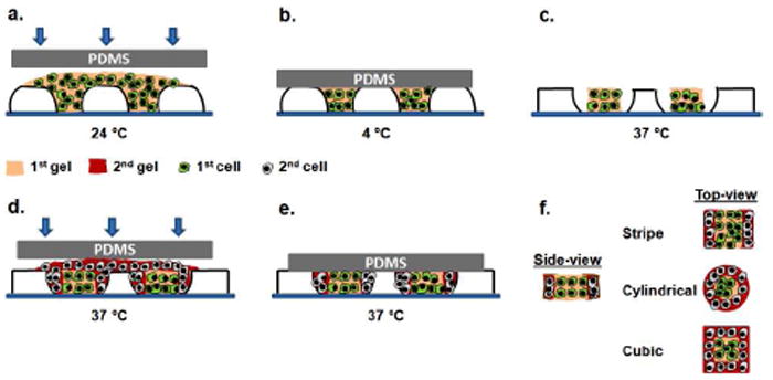
Schematic diagram of sequential patterning of hydrogel microstructures with responsive micromolds. Either fluorescent microbeads or cells (3T3 fibroblasts, HepG2 cells and HUVECs) were encapsulated within agarose microgels during fabrication process. a) The first gel precursor with encapsulated cells or microbeads was put on a responsive micromold and molded with a PDMS slab at 24 °C. b) 15 min incubation at 4 °C for crosslinking the first gel. c) 30 min incubation at 37 °C to allow responsive micromolds to shrink. d) The second gel precursor with encapsulated cells or microbeads was put on a responsive micromold and molded with a PDMS slab at 37 °C. e) 30 min incubation at 37 °C for crosslinking the second gel. f) Side and top views of resulting hydrogel microstructures.