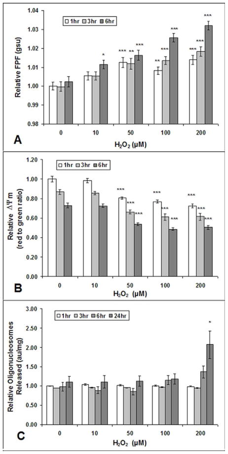
Fig. 1. FPF, Δψm, and apoptosis in cultured human RPE cells after exposure to H2O2.
Increased FPF (A) and reduced ΔΨm (B) at 1, 3, and 6 hours after cultured human RPE cell exposure to various H2O2 concentrations. Human RPE cells did not show increased levels of apoptosis (released mono- and oligonucleosomes) measured by Cell Detection ELISA, until 24 hours after exposure to 200 μM H2O2 (C). Values are mean (standard error). *P<.05, ***P<.001, compared with corresponding control (0 μM H2O2). Each experiment was conducted on 3 different cell lines, n=9.

