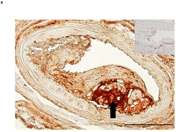
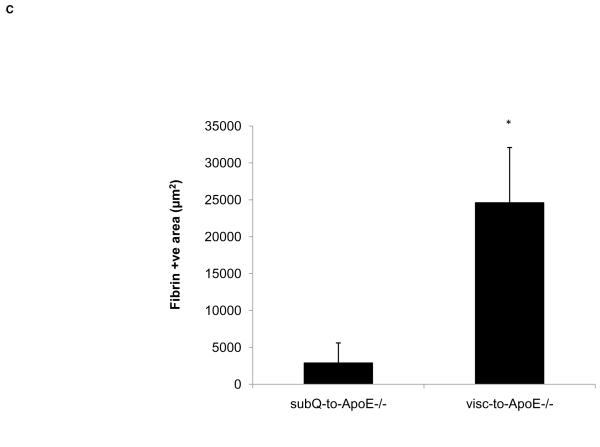
Figure 4. ApoE−/− mice transplanted with visceral fat display complicated lesions.
A) Cross section of carotid artery stained with fibrin(ogen) antibody from ApoE−/− mice transplanted with A) subcutaneous and B) visceral fat. Inset shows negative control without primary antibody. Arrow points to necrotic core of the lesion. Scale bar = 100 μm, magnification 40x. C) Fibrin-positive area in lesions in operated mice. *p<0.05.


