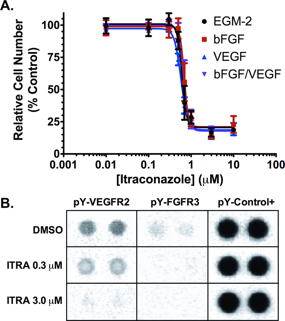
Figure 1. Inhibition of proliferation and receptor tyrosine kinase phosphorylation in stimulated HUVEC cultures.
(A) Itraconazole inhibits HUVEC proliferation in a dose dependent manner in cultures stimulated by supplementation with EGM-2 (black), 12 ng/ml bFGF (red), 10 ng/ml VEGF (blue), and 12 ng/ml bFGF with 10 ng/ml VEGF (purple). Mean relative cell number was evaluated by MTS assay and are reported as mean percent of vehicle treated cell proliferation ± SD. (B) Cell lysates from EGM-2 stimulated HUVEC treated with 0.3 µM or 3.0 µM itraconazole, or with vehicle control for 24 h were hybridized to an RTK array and probed with anti-phospho-tyrosine-HRP antibody followed by chemiluminescent detection. Each RTK is spotted in duplicate with signal normalized to phospho-tyrosine internal controls.

