
Figure 5.
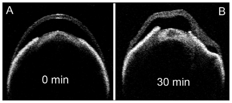
Time-lapse images of a representative single eye exposed to conventional fixative. The Visante OCT was used for ex vivo imaging of the whole eye immediately after placing in 2.5% glutaraldehyde fixative solution (A) and after 30 minutes (B). Distinct stromal swelling and distortion are evident by 30 minutes.