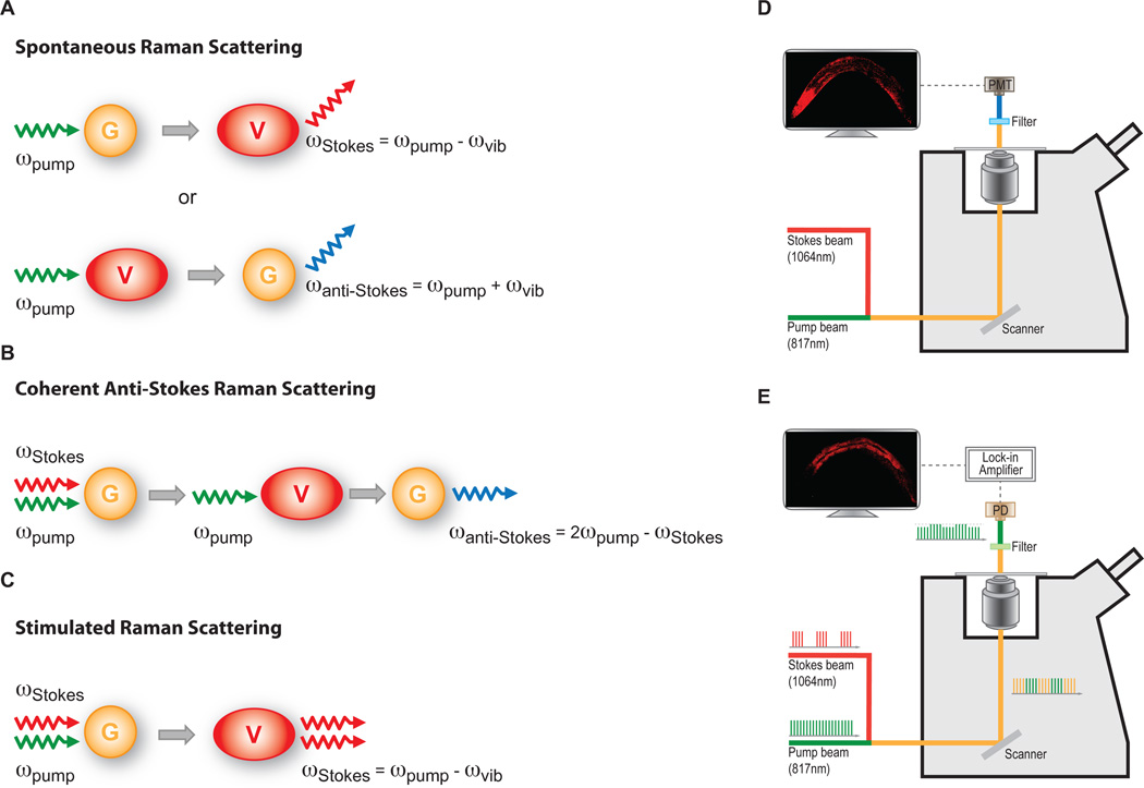
Figure 1. Principle and diagram of CARS and SRS microscopy systems.
A. Spontaneous Raman scattering. Incident photons interacting with molecules normally scatter elastically. However, a very small fraction of photons are inelastically scattered. When an incident photon (pump photons) interacts with a molecule carrying specific chemical bonds in the vibrational ground state (G), the inelastic scattering photon (Stokes photon) will have lower energy than the incident ones. The energy difference between the pump and Stokes photons is equal to the vibrational energy of the chemical bond, ϖpump - ϖStokes = ϖvib. In contrast, a scattering photon with a higher energy (Anti-Stokes photons) will generate, by interacting with the chemical bonds in an excited vibrational state (V), ϖanti-Stokes - ϖpump= ϖvib.
B. Coherent anti-Stokes Raman scattering. This multiphoton process is achieved by using higher energy “pump” photons and lower energy “Stokes” photons, with energy difference being resonant with the vibrational frequency of specific chemical bonds in a molecule. The non-linear interaction between pump and Stokes photons will excite the vibration coherence of the chemical bonds in the molecule. This excited vibration coherence will further interact with a second pump photon, resulting in the coherent emission of an anti-Stokes photon that is more energetic (ϖanti-Stokes = 2ϖpump - ϖStokes = ϖpump + ϖvib). Unlike spontaneous Raman scattering, where inelastic photons scatters in all directions, the anti-Stokes light in CARS emits in a certain specific direction.
C. Stimulated Raman scattering. When the energy difference between pump and Stokes photons resonant with the vibrational frequency of a type of chemical bonds in a molecule. The non-linear interaction between two photons stimulates the chemical bonds into excited vibrational state. The energy transfer from the pump beam to the Stokes beam accompanies this, vibrational excitation, resulting in the disappearance of one pump photon and the creation of a Stokes photon.
D. Diagram of CARS microscope. The Stokes laser beam has a fixed wavelength at 1064nm, while the pump laser beam is tunable. Pump beam at 817nm for lipid imaging is used an example here. The combined pump and Stokes laser beams is scanned over the sample by a XY scanner. CARS signal is collected using a red-sensitive photomultipier tube (PMT). In front of the PMT, filters are used to block the pump and Stokes beams and any induced two-photon fluorescence.
E. Diagram of SRS microscope. The laser set up is very similar to CARS, except that the pump laser beam is modulated at a high frequency (~10MHz). The filter is used to block the Stoke beam completely. The transmitted pump beam containing SRL signals is detected by a photodiode (PD). An electronic device, Lock-in Amplifier, is used to demodulate the SRL signals carried on the pump beam.

