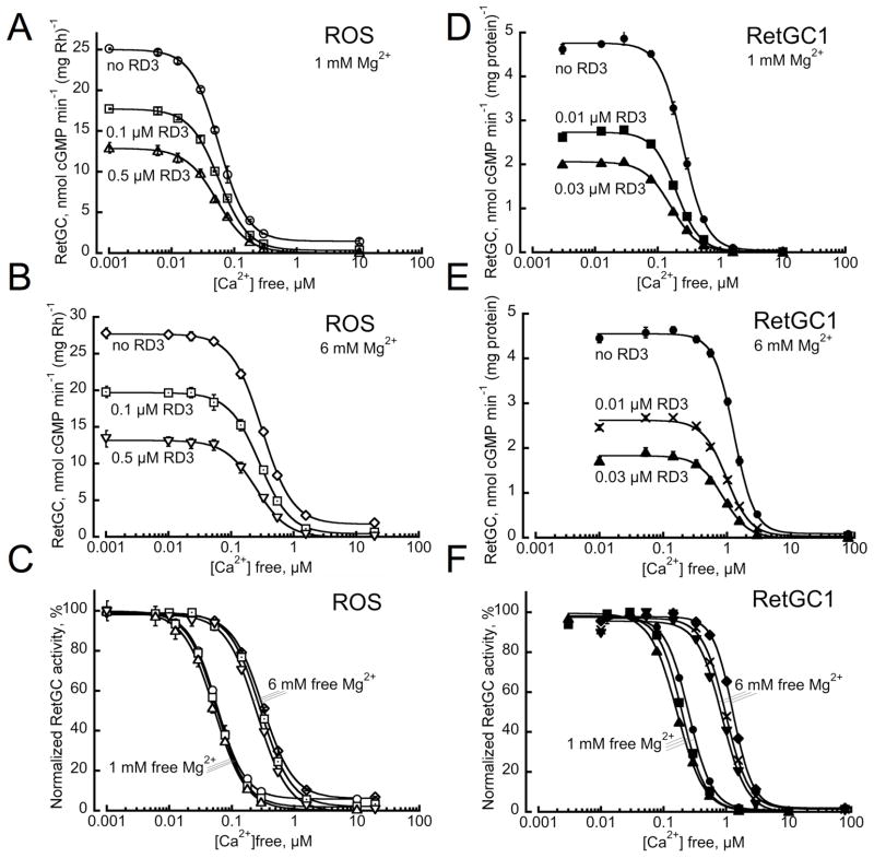
Fig. 3.
RetGC activation by GCAP suppressed by RD3 retains near normal sensitivity to Ca2+and Mg2+. A–C. ROS fraction isolated from C57B6 mouse retinas was assayed for RetGC activity (open symbols) at various free Ca2+ concentrations and either 1 mM (A) or 6 mM (B) free Mg2+ concentrations in the presence or in the absence of 0.1 μM or 0.5 μM recombinant RD3 as indicated; in panel C, the activities normalized to the maximal RetGC activity in each assay series are shown as a function of free Ca2+ concentrations. D–F. Membrane fraction from HEK 293 cells expressing human RetGC1 (filled symbols) was reconstituted with 5 μM recombinant GCAP1 in the absence or in the presence of 10 nM and 30 nM RD3 as indicated and assayed as described under Experimental Procedures.

