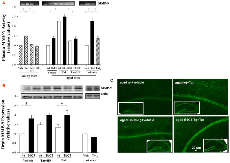
Figure 3. Tat-induced MMP-9 expression is potentiated in B6C3-Tg mice.
Mice were injected with Tat, Tathi, or hydroxyfasudil (HF) as in Figure 1 and analyses were performed 24 h post Tat injection. (A) Plasma MMP-9 activity was determined by zymography. (B) Expression of MMP-9 protein in the brain tissue was assessed by Western blotting. Data are mean ±SEM, n= 4–5; *, p< 0.05; †, p< 0.01. (C) In situ zymography was performed to visualize gelatinase activity in the hippocampal sections of aged B6C3-Tg and control mice. Images are representative from 4 experiments.

