
Fig. 2.
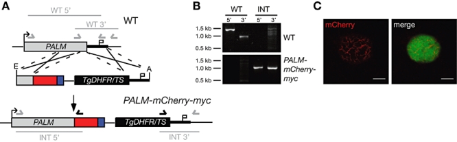
Live cell imaging of PALM in infected hepatoma cells. A. Generation of PALM-mCherry-myc parasites. The PbPALM genomic locus was targeted with a replacement plasmid containing the C-terminal PALM fragment (grey) fused in-frame to the mCherry coding sequence (red), and a quadruple c-myc tag (blue) and followed by the 3′UTR of PbDHFS/FPGS. In addition, the targeting plasmid contains the PbDHFR/TS positive selectable marker (black box), and a fragment of the PALM 3′UTR. Upon a double cross-over event, the targeting plasmid is expected to replace the endogenous PALM ORF with a C-terminally tagged PALM fusion protein. Arrows and bars indicate specific primers and PCR fragments respectively. B. Genotyping of the PALM-mCherry-myc parasite line. Using integration-specific primer combinations (A), the successful replacement event was verified. Absence of the WT signal from PALM-mCherry-myc parasites confirmed the purity of the clonal population. C. Hepatoma cells were infected with PALM-mCherry-myc sporozoites. PALM expression was visualized in late liver stages 2 days after infection. Note the branched structure of the mCherry signal. Bars, 10 µm.